19 October 2018
Researchfunds for unique 3D imagery
The Swedish Foundation for Strategic Research (SSF) has allocated 6.8 million to researchers at Luleå University of Technology to develop a new imaging method that can be of great importance to future research and innovation, in cancer detection, material science, environmental research and industry.
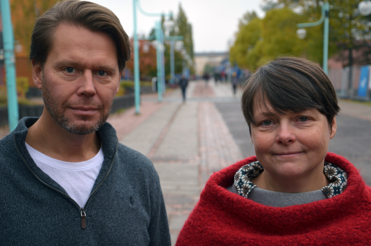
– We hope that we will be able to develop an imaging method that enables us, among many applications, to detect cancer at an earlier stage and in a more cautious way.With this method we do not have to alter the tissue or use hazardous radiation to measure it, Kerstin Ramser says, Professor of Experimental Mechanics at Luleå University of Technology.
3D imaging method
The goal of the researchers in Experimental Mechanics at Luleå University of Technology, is a new 3D imaging method, termed InFeRa. For example, in medical research, there is a great need to be able to take three dimensional (3D) in vivo images of specific species and to minimize the impact on the sample. Today, a sample has to be taken out of the body or from a trial set and colored in, which often destroys or modifies the sample. InFeRa combines three methods into a compact device with a software for recording, analyzing and displaying 3D images.
– We will build a completely new imaging method based on our previous research in the field, so the method is already under development by our research team," Mikael Sjödahl, says Professor of Experimental Mechanics at Luleå University of Technology.
InFeRa combines spatially gated Interferometric imaging (InFe), with stimulated Raman scattering (Ra) that provides information about a substance's concentration and location as well as of biochemical processes. A spatial light modulator provides a programmable interference pattern that can be used for controlled 3D scanning through the sample. InFeRa will be able to reduce the need for separate, costly, time-consuming chemical and enzymatic in vitro methods.
Collaboration with Biochemical Process Engineering
Even in industry and environmental research, you want to be able to take 3D pictures in situ to find out where, how and why some substances are formed. There is a need to be able to study biochemical processes or to see how a substance affects the properties of a material. Initially, the researchers will test InFeRa on biofilms (films of microorganisms on surfaces) in bioelectronic systems (BES) in cooperation with researchers in Biochemical Process Engineering at Luleå University of Technology. The goal is to in situ study energy transfer, molecular signaling, activities, reactions to environmental change, growth and aging of biofilms. BES are an important technology for future energy constraints.
– The goal is to show a 3D structure of specific species, such as, in this case, a protein, where it is, how it is produced and react to environmental change and how specific substances affect different materials, Mikael Sjödahl, says.
– For our part, we hope that the new imaging method InFeRa can help to improve the efficiency of production of electricity in microbial fuel cells and the production of chemicals from CO2 in so-called microbial electrolytic cells. This technique, InFeRa, is particularly attractive as it can scan surfaces even in depth, Magnus Sjöblom, says, researcher in biochemical process engineering at Luleå University of Technology.

Published:
Updated: