Laboratories and equipment at LUMIA
Within LUMIA, a large variety of solid materials are analyzed. Characterization is done both in terms of imaging and chemical composition, usually on a micro- to nanometer scale.
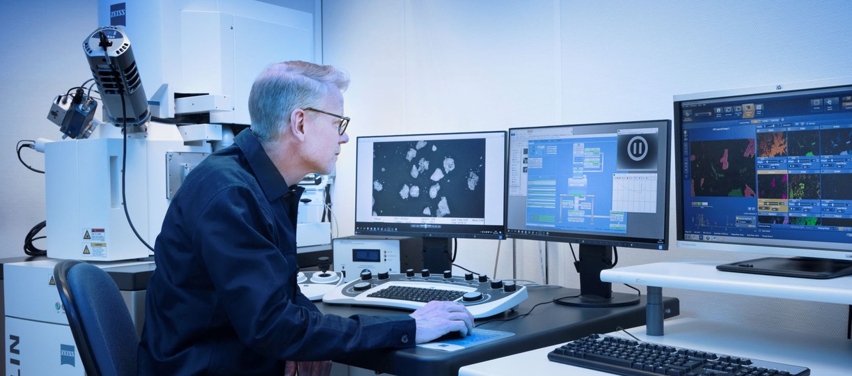
Scanning Electron Microscopy - Zeiss Merlin
Zeiss Merlin FEG-SEM with EDS/WDS. The microscope has an image resolution of about 0.8 nm and is equipped with:
- dual BSE detectors
- dual SE detectors (standard and in-lens)
- EDS (energy dispersive spectrometry)
- WDS (wavelength dispersive spectrometry)
For EDS/WDS analysis, the Aztec/Inca analysis platforms from Oxford Instruments are used. Opportunities for automated mineralogy are available via Aztec Feature and Large Area Mapping.
Responsible: Mathis Warlo
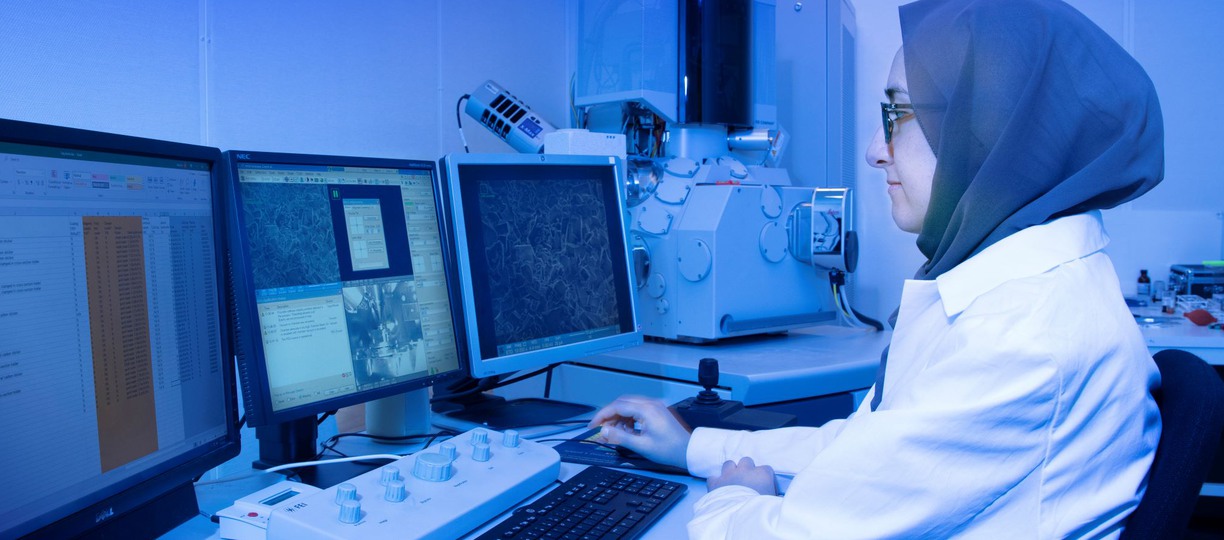
Scanning electron microscopy - FEI Magellan
FEI Magellan 400 FEG-SEM (extreme high resolution SEM). The microscope has an image resolution of about 0.5 nm (0.5-30 kV) and is equipped with:
- ETD: Everhart-Thornley detector
- TLD: in-lens detector (extreme high resolution)
- vCD: Backscattered electron detector (BSD).
- STEM: Scanning Transmission Electron Microscopy (STEM) detector
- EDS: energy dispersive spectroscopy detector, X-Max 80 mm2 SDD, Oxford Instruments.
- EBSD: HKLNordlysN detector for electron backscatter diffraction (Oxford Instruments).
Responsible: Liang Yu
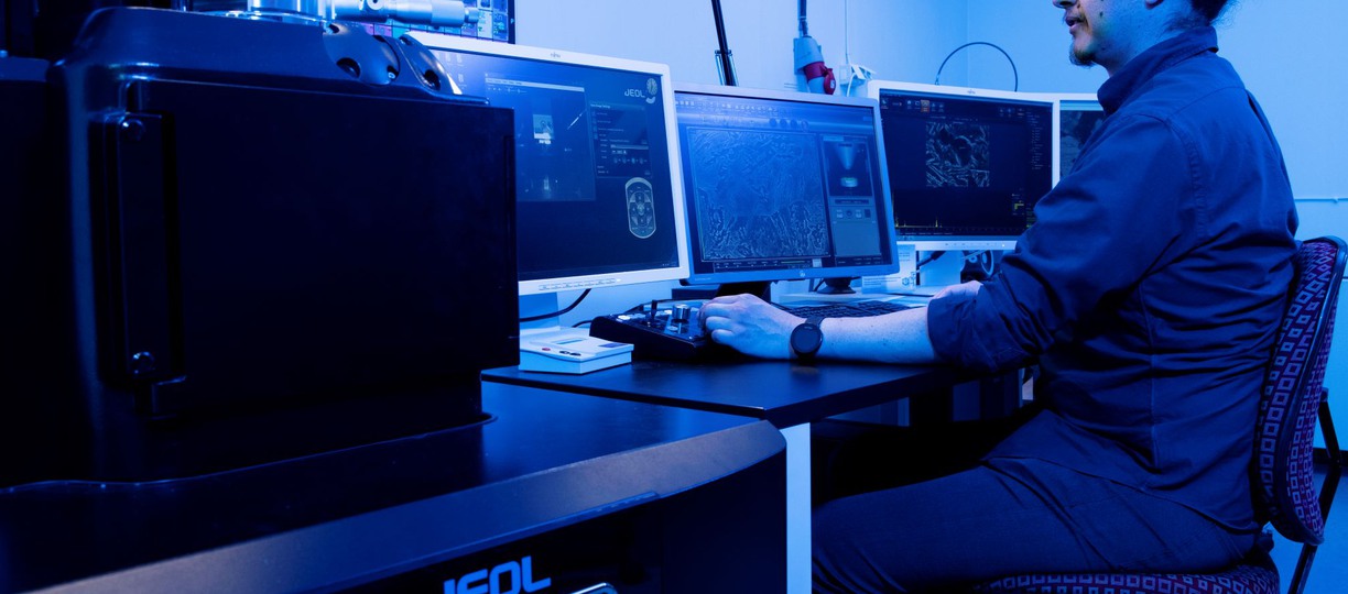
Scanning Electron Microscopy - Jeol JSM-IT300
Jeol JSM-IT300 Low and high vacuum SEM with EDS and EBSD. Image resolution of approximately 10 nm and is equipped with:
- Ability to analyze in low vacuum
- Large chamber for large samples or details
- BSE detector
- SEI detector
- EDS: X-Max 80 from Oxford Systems
- EBSD: Nordlys Max3 from Oxford Systems
For EDS and EBSD, Aztec and Channel5 from Oxford Systems are used.
Responsible: Erik Nilsson
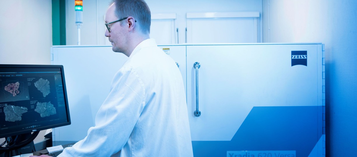
X-ray tomography - Zeiss Xradia 620 Versa
ZeissXradia 620 Versa (Carl Zeiss Microscopy)
- 3D imaging of internal microstructure
- Max spatial resolution: 0.5 μm (Min voxel size: 40 nm)
- Field of view (FOV): 0.1-50 mm (Sample size: 0.1-200 mm)
- Absorption and phase contrast imaging
- In-situ loading module: Deben CT5000TEC (5 kN compression/tension, temperature -20/+160°C)
- 3D crystallographic imaging with LabDCT (lab-based Diffraction Contrast Tomography)
- Quantitative analysis of structure (size distribution, shape, volume fraction of particles/fibers/cells/defects etc)
- Quantitative analysis of deformation (3D analysis of displacement and strain with DVC).
LUMIA can handle most of the sample production and sample preparation for analysis and is also used to handle unconventional sample types.
Responsible: Henrik Lycksam
Updated:
Page author: Contact us